
A 46-year-old man was admitted to the hospital with COVID-19. On arrival he was moderately dyspneic, with an oxygen saturation of 89% on room air. He was started immediately on supplemental oxygen at 3 L/min via nasal cannula1. He was also started on dexamethasone and remdesivir2. Another patient was brought in with him: his 72-year-old mother. She had developed a fever and a cough several days after her son did, and she too was diagnosed with COVID-19 — a household transmission. Because she had long-standing diabetes, she was at high risk of severe disease. She was also admitted and started on supplemental oxygen at 1 L/min via nasal cannula in a different isolation room.
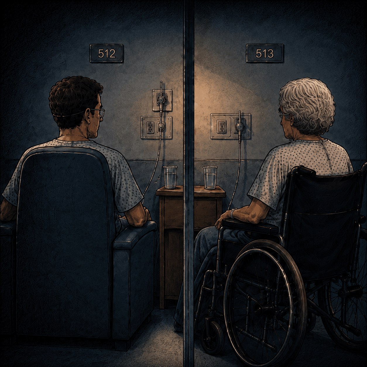
The son was stable, but his mother deteriorated rapidly. A chest X-ray on the third day showed bilateral diffuse infiltrates3. She required 10 L/min to maintain her saturation level. Our team held a conference with the critical care team, where we discussed whether she needed to be transferred to the intensive care unit (ICU). The pandemic was raging at the time. The ICU was already close to full, and there were other patients on the ward with severe pneumonia, or expected to deteriorate soon. After careful discussion and consideration of her wishes, we decided to continue her care on the ward.
The next day, I performed an electrocardiogram (ECG)4 on her son. The tracing showed deep, biphasic and inverted T waves in leads V2 and V35. This is a classic sign of Wellens syndrome, which indicates a critical narrowing of the left anterior descending artery, the main vessel supplying blood to the front of the heart. The artery could close at any moment, causing a massive heart attack that very few people survive. My first thought was, "Oh, this is exactly what I saw in that Images in Clinical Medicine piece in the New England Journal of Medicine (NEJM)6." I knew what I was looking at. I felt obligated to tell him immediately, so that he could be evaluated by a cardiologist and treated before it was too late.
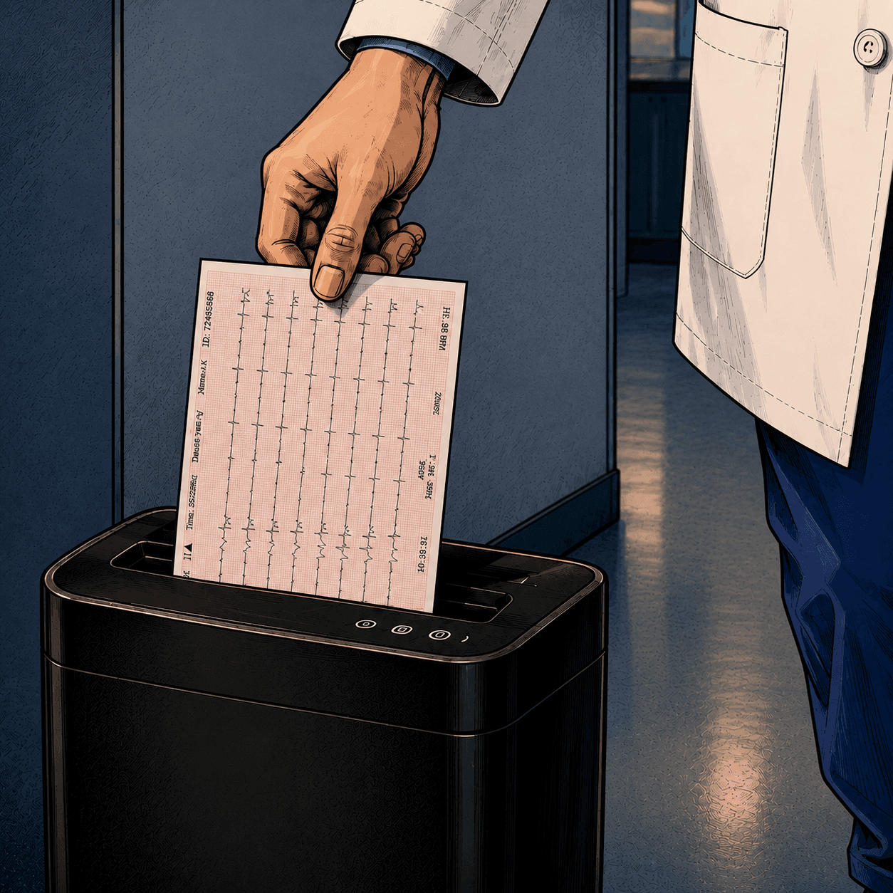
I printed the tracing and went up to the ward to tell him about the finding. I put on the full set of personal protective equipment: a gown, gloves, an N95 mask, and a face shield. The printout went into a plastic folder so it would not be contaminated. I knocked on the door and opened it. But he was not there. He was not supposed to be outside the room for infection control reasons. Where could he be?
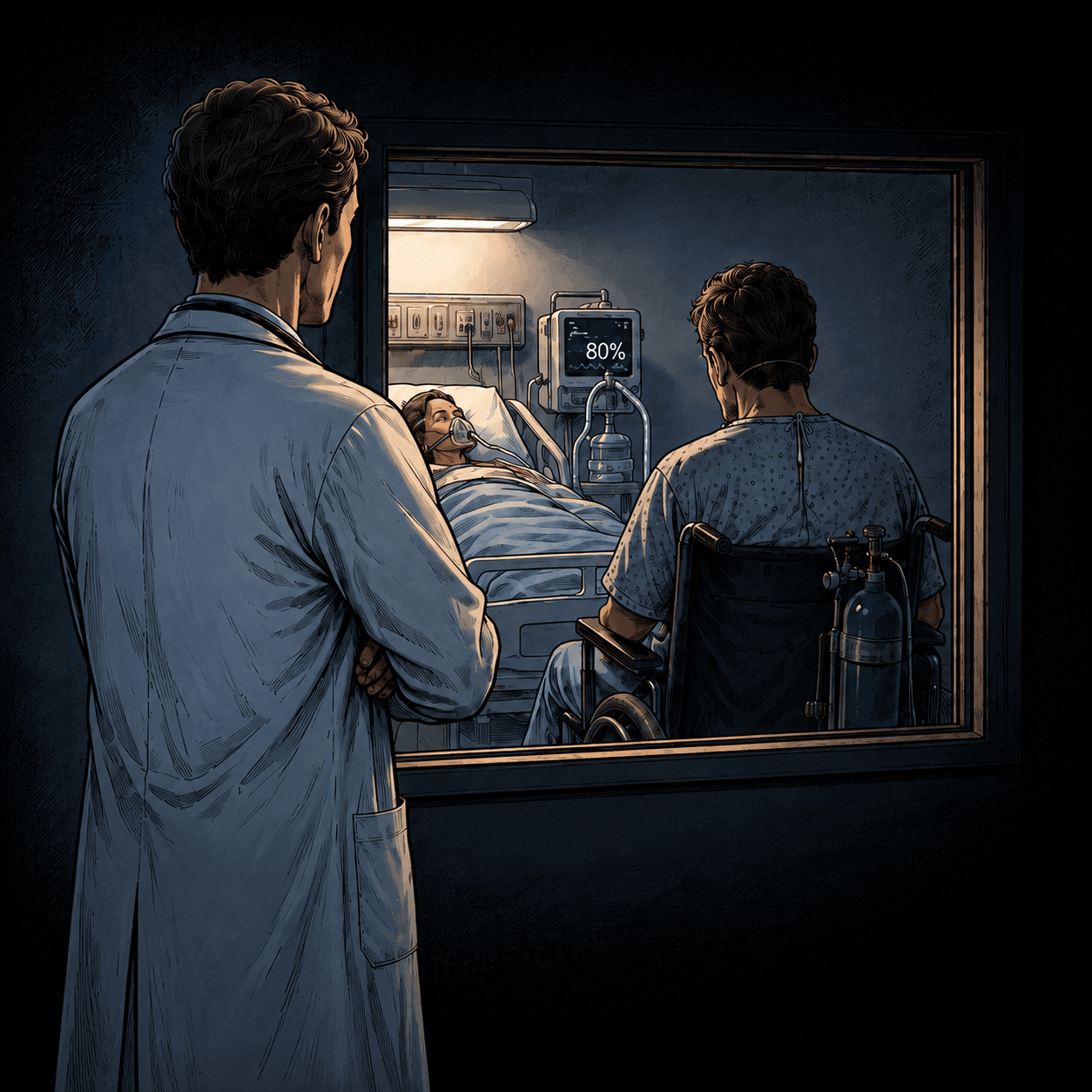
The nurse told me he was in his mother's room. Her condition had worsened rapidly. She could barely hold her oxygen saturation above 90% at the maximum flow rate. Our team had discussed her case again that morning and had listened to her wishes and her family's. Her COVID-19 pneumonia had reached the point of no return. We had to move to palliative care. The nurses had arranged a final moment together for her and her son. The two of them had lived together at home for decades until they were admitted. He sat beside her. No other family members were allowed to see her, because of the risk of transmission. He might have been there alone, facing the remorse of having infected her.
He could not have cared less about the narrowing in his coronary artery, or the risk of a heart attack, or any of it at that moment. Looking back now, I am really grateful that I stopped before I actually blurted out the ECG finding to him. I shredded the ECG printout at the ward nurses' station and left them alone. This was the moment I realized I was becoming a physician-robot. Analyzing a patient's condition is of course part of a physician's duty. What I did was not wrong. But the problem was that I had no idea what must have been going on in his mind until I saw him sitting there with his mother. I had been so focused on the data that I had completely lost sight of what really mattered to him.
Every physician is expected to analyze patient data, and the volume of it is often beyond what a single human mind can hold. Every day, new patients come and go with acute conditions, frequently on top of several chronic ones. For each of them you are expected not to make a single mistake as you review an ECG, dozens of laboratory results, hundreds of CT slices that might hide something subtle and unexpected such as an incidental tumor, and walls of text in the electronic health record documenting a medical history that often goes back many years. Day after day, faced with this volume of information, you are programmed to process and analyze patient data as accurately as possible — pretty much like training a machine learning model.
You don't have to be a machine. We now have machines that can help you do the job. AI has reached a point where it can understand human text and images, and other modalities such as audio and video, well enough to carry out everyday and professional work for people: writing, coding, designing. What is overwhelming about clinical medicine is the sheer quantity of data a physician is now expected to take in. Processing and analyzing data is precisely what AI is built for. It can already read research papers far faster than any human. Perhaps it is time for us to consider seriously whether AI should be helping physicians carry that load. I moved into this field so that physicians do not have to forget the best part of medicine.
Footnotes
-
Supplemental oxygen: Oxygen given to patients whose blood oxygen levels are low. 1 to 3 L/min is generally considered low-flow therapy, while 10 L/min is at the high end of what a mask can deliver. ↩
-
Dexamethasone and remdesivir: Medications used in the treatment of COVID-19. This combination was the standard of care for hospitalized patients with COVID-19 pneumonia at that time. ↩
-
Infiltrates: A term used in radiology for abnormal shadows in the lung fields, often indicating pneumonia or other lung disease. ↩
-
ECG (electrocardiogram): A test that records the electrical activity of the heart, used to detect cardiac problems. It is one of the most common diagnostic tools in medicine. ↩
-
T waves in leads V2 and V3: The T wave is the deflection seen just after each contraction of the heart. V2 and V3 are two of the twelve views the machine records, and they look at the front wall of the heart. ↩
-
New England Journal of Medicine: A very famous journal, and one of the most prestigious in medicine. Essentially every physician knows it. ↩